Biology
You were taught:
Dogs see the world in black and white.
Now we know:
Dogs see colors, but their spectrum is limited compared to humans. They see shades of blue and yellow but cannot distinguish red and green.
Disproven1989
Read more →Blood is always red. Deoxygenated blood is dark red, not blue. Veins appear blue through skin due to light scattering and tissue absorption.
Hold your wrist to the light and the veins trace blue-green rivers beneath the skin. The color seems self-explanatory. Arteries carry oxygenated blood, which is red; veins carry deoxygenated blood; deoxygenated blood must therefore be blue. The argument is neat, internally consistent, and wrong at its premise.
Blood is always red. Oxygenated blood is bright scarlet, hemoglobin bound to oxygen reflects the shorter wavelengths of the visible spectrum. Deoxygenated blood, hemoglobin with its oxygen removed, is a darker, more burgundy red. But it is not blue. It has never been blue. Any blood draw, any surgical procedure, any observation of venous blood outside the body confirms this immediately: the blood in the tube is dark red, not blue or violet or anything approaching the color one sees through intact skin. The difference between arterial and venous blood is a difference of shade, not of color family.
The explanation for the visual phenomenon was worked out rigorously by Alwin Kienle and colleagues at the Institute for Laser Technologies in Medicine in Ulm, Germany. Their 1996 paper in Applied Optics, "Why do veins appear blue? A new look at an old question," used both experimental CCD camera measurements and Monte Carlo light-transport simulations to model how light interacts with skin, subcutaneous fat, and blood vessels of varying diameter and depth. The findings were precise: the apparent color of a vein as seen through skin is not the color of the blood inside it. It is the product of differential light absorption and scattering by the surrounding tissue.
Skin absorbs different wavelengths of light at different rates. Red and near-infrared wavelengths penetrate deeply into tissue and are heavily absorbed by both oxygenated and deoxygenated hemoglobin. Blue wavelengths are absorbed more strongly by the superficial layers of skin. In the specific geometry of a blood vessel beneath several millimeters of tissue, the optical properties of the surrounding layers interact with the vessel's absorption spectrum in a way that causes the region above the vein to appear blue-green to the eye, even though the vessel is full of dark red blood. The exact hue depends on vessel depth, diameter, surrounding fat content, and skin tone; very deep or very narrow veins may appear no different from surrounding tissue.
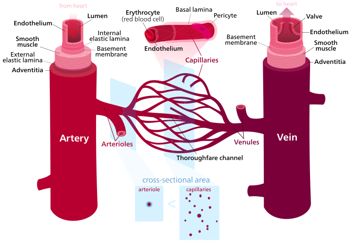
The biological intuition that blue equals deoxygenated had a certain cultural reinforcement: medical diagrams consistently colored arteries red and veins blue as a convention for distinguishing the two systems, and the convention was frequently misread as anatomical fact. Textbook illustrations, classroom posters, and anatomical models all used blue for veins, not because anyone had measured the color of venous blood and found it blue, but because blue was the most visually legible contrast to arterial red. Generations of students encountered the diagram before they encountered a physiology explanation, and the diagram's logic appeared to confirm what one could see in the mirror. The self-reinforcing loop was strong enough to survive intact into an era when the actual optics had long been worked out. The myth persisted not through neglect of the evidence but through a diagram that arrived first and a question that almost nobody thought to ask.
Dogs see the world in black and white.
Dogs see colors, but their spectrum is limited compared to humans. They see shades of blue and yellow but cannot distinguish red and green.
Bulls are enraged by the color red.
Bulls are colorblind to red. They react to the movement of the matador's cape, not its color. The cape is red for tradition and to hide blood stains.
Watching television occasionally is harmless to eyesight, and the radiation from TV sets is negligible.
Early color CRT televisions (particularly GE's 1967 recall) did emit low levels of X-ray radiation at close range. Modern concerns shifted to non-ionizing blue light and screen time effects on vision development in children.
Camels store water in their humps to survive in the desert.
Camels store fat in their humps, not water. Water conservation comes from efficient kidneys, concentrated urine, and tolerance to dehydration.