Humans have 206 bones as adults, and this number is fixed and universal.
The number of bones varies individually. Many adults have extra ribs, sesamoid bones, or fused/split bones. The exact count ranges from 206 to over 270 in infants whose bones haven't fused.
What changed?
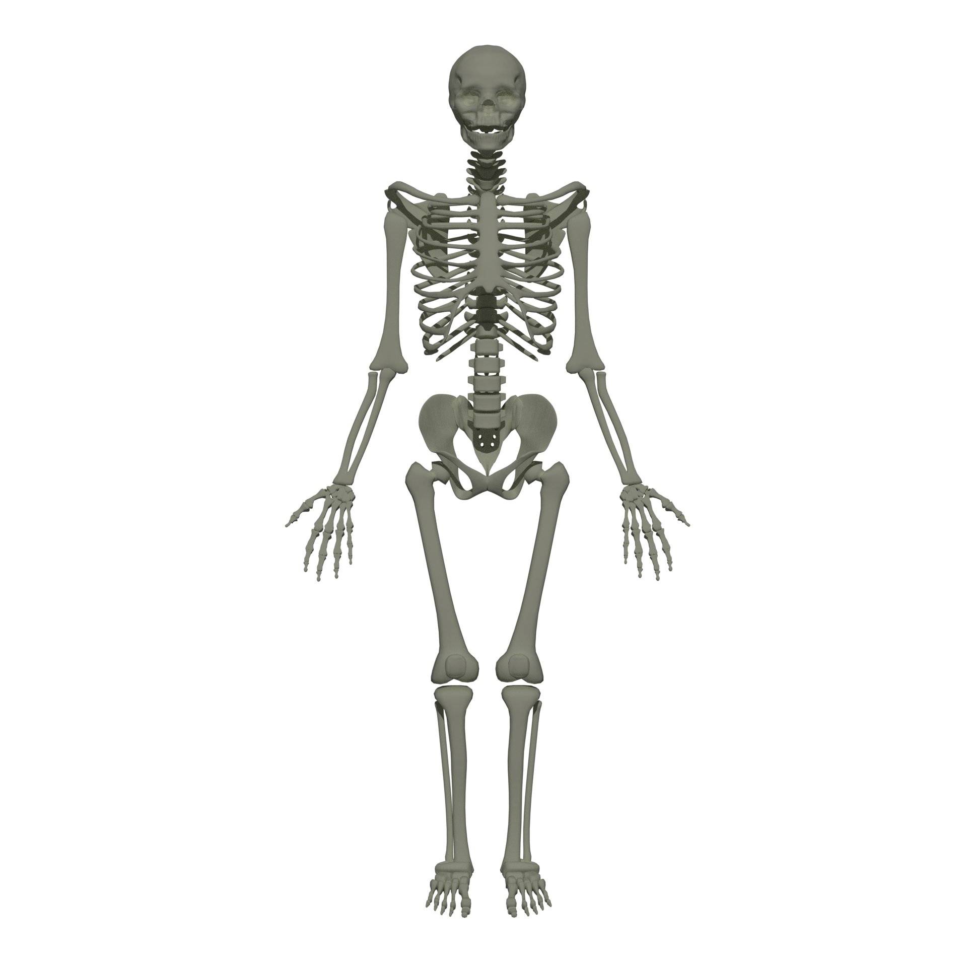
Anatomy textbooks have told generations of students the same number: 206. That is how many bones an adult human body contains. The figure appears in elementary school science lessons, in medical school curricula, in crossword puzzle clues, and in popular science writing. It is stated as a fixed fact about the human body, as determinate as the number of chambers in the heart.
The number is not fixed. It never was.
The figure 206 represents an average, more precisely, the count that emerges when an anatomist surveys a typical adult skeleton and tallies the bones that are usually present. In practice, the human skeleton varies considerably from one individual to another, and those variations are not anomalies or pathologies but normal expressions of biological variation in a species with substantial genetic and developmental flexibility.
The most common source of variation is sesamoid bones. These are small, rounded bones that develop within tendons, typically near joints subject to high mechanical stress. The patella, the kneecap, is the largest sesamoid in the body and is included in the standard count of 206. But many people develop additional sesamoid bones in the hands and feet, and a small bone behind the knee called the fabella is present in some individuals and absent in others. A 2019 systematic review published in the Journal of Anatomy by Michael Berthaume and colleagues analyzed skeletal data from more than 21,000 knee studies spanning 150 years and found that fabella prevalence had increased from 11 percent of the population in 1918 to 39 percent by 2018, a more than threefold increase, apparently driven by rising body mass and increased mechanical loading. The bone can appear or not appear across a human lifetime, depending partly on genetics and partly on physical forces.
Sutural bones, also called Wormian bones after the Danish anatomist Ole Worm, are small, extra bones that develop along the suture lines of the skull. They form when the fibrous sutures between skull bones ossify in irregular patterns, producing small additional islands of bone within what would typically be a single plate. Most people have a few, or none. Some individuals have dozens. They are not associated with disease or dysfunction; they are simply variation.
The infant skeleton complicates the count further. Newborns have somewhere between 270 and 300 bone elements at birth, because many structures that later fuse into single bones, the sacrum, the sternum, the hip bones, exist as separate ossification centers in infancy and early childhood. Fusion proceeds throughout childhood and into early adulthood; the clavicle, the last bone in the body to fully fuse, typically completes the process between the ages of 25 and 35. The adult count of 206 therefore reflects not a stable feature of the skeleton but the typical endpoint of a decades-long consolidation process that varies in timing among individuals.
Some adults never complete certain fusions. The os trigonum, a small bone at the back of the ankle, typically fuses to the talus during adolescence; in a small percentage of the population, it remains separate into adulthood. The metopic suture of the frontal bone, which typically fuses in early childhood, producing a single frontal bone from two halves, remains open in a minority of adults.
The teaching of 206 as a universal, fixed number reflected pedagogical convenience rather than biological reality. An anatomy lesson that presents variation requires more nuance than a lesson that presents a single number. But the convenience came at the cost of accuracy, and it encouraged a view of the human body as more standardized than it actually is. Human skeletons differ. The number of bones is one of the ways they do.